- This topic is empty.
-
AuthorPosts
-
2026-10-10 at 6:02 pm #89618
Recognizing the Clinical Picture of Male Hypogonadism
Male hypogonadism can present through a wide range of overlapping symptoms, including low libido, erectile dysfunction, gynecomastia, reduced muscle mass, fatigue, depression, osteoporosis that is not explained by aging, delayed puberty, and testicular abnormalities. Because these signs can also reflect metabolic, psychological, or unrelated endocrine conditions, clinicians rely on laboratory evaluation of the hypothalamic-pituitary-gonadal axis to confirm androgen deficiency and determine its origin. Testosterone, LH (Luteinizing Hormone), and FSH (Follicle-Stimulating Hormone) form the core panel used for this purpose, as each marker reflects a different point along the reproductive endocrine pathway.
Testosterone as the Primary Androgen Status Signal
Testosterone is described as an androgen marker for gonadal and adrenal androgen status, and its differentiated value lies in supporting the assessment of androgen excess or deficiency in both female and male reproductive and metabolic conditions. In men, decreased testosterone is associated with hypogonadism and aging-related androgen decline, while elevated testosterone in men is more often linked to testicular tumors or steroid abuse rather than hypogonadism itself. Within hypogonadism-specific interpretation, testosterone is markedly decreased (↓↓) in Primary Testicular Failure and Klinefelter Syndrome, and decreased (↓) in Hypogonadotropic Hypogonadism affecting both females and males. On its own, however, a low testosterone result does not indicate whether the defect originates in the testes or in the hypothalamic-pituitary axis above it — this is where LH and FSH become essential.
LH and FSH as Gonadotropin Signals
LH is positioned as a pituitary gonadotropin marker for ovarian and testicular function, with a differentiated value centered on distinguishing primary gonadal failure from central hypothalamic or pituitary causes when interpreted alongside FSH and sex steroids. In men, elevated LH is associated with primary testicular failure and Klinefelter syndrome, while decreased LH points toward hypothalamic or pituitary dysfunction, such as Kallmann syndrome.
FSH plays a parallel role as a gonadotropin reserve signal, also used to evaluate testicular function. Elevated FSH in men is associated with primary testicular failure, while decreased FSH is linked to hypothalamic/pituitary disorders and hyperprolactinemia. Because LH and FSH respond to signals from the testes through feedback loops, their behavior relative to testosterone reveals where in the axis the dysfunction is occurring. This is the central logic behind evaluating all three analytes together rather than testosterone alone.
Differentiating the Underlying Cause
The disease-specific interpretation data highlights three distinct patterns relevant to suspected male hypogonadism:
Primary Testicular Failure shows LH elevated (↑←) and FSH markedly elevated (↑↑), alongside markedly decreased testosterone (↓↓). The clinical interpretation note indicates that primary testicular failure should be confirmed with testicular ultrasound.
Klinefelter Syndrome shows both FSH and LH markedly elevated (↑↑) together with markedly decreased testosterone (↓↓). The clinical interpretation note specifies that this pattern — FSH/LH markedly elevated plus decreased testosterone — should be confirmed with karyotype testing for 47,XXY.
Hypogonadotropic Hypogonadism shows the opposite gonadotropin pattern: both LH and FSH are markedly decreased (↓↓), while testosterone is decreased (↓) in both females and males. The clinical interpretation note specifies that pituitary lesions should be excluded when this pattern is identified.
In each case, testosterone alone establishes that androgen deficiency is present, while the relative behavior of LH and FSH indicates whether the cause is primary (testicular) or central (hypothalamic-pituitary), and points toward the specific confirmatory step — testicular ultrasound, karyotype analysis, or pituitary lesion exclusion — that should follow.
Supporting Interpretation Within a Broader Endocrine Context
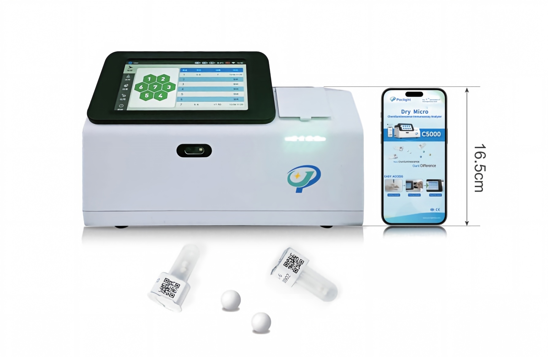
Reliable interpretation of Testosterone, LH, and FSH does not occur in isolation. The Sex Hormones Clinical Knowledge Graph reflects a multi-tier endocrine pathway assessment designed to support separation of central and peripheral dysfunction by evaluating brain centers, pituitary regulators, adrenal glands, gonads, and placental signals together. As part of Poclight’s 9-analyte Sex Hormones profile — which also includes β-hCG, AMH, Cortisol, Progesterone, E2 (Estradiol), and Prolactin (PRL) — Testosterone, LH, and FSH results can be considered alongside related markers such as Prolactin, since hyperprolactinemia is itself associated with decreased LH and FSH and can mimic or contribute to a hypogonadal presentation. This layered structure is intended to support clinical evaluation of reproductive development, fertility, libido, bone health, metabolic balance, menstrual regulation, and spermatogenesis across conditions that a single marker cannot fully explain.
This approach to androgen deficiency evaluation is consistent with the broader clinical literature on testosterone therapy in men with hypogonadism, including guidance published in the Journal of Clinical Endocrinology & Metabolism (Bhasin et al., 2018), which underscores the importance of confirming androgen deficiency biochemically before pursuing further diagnostic workup.
A Structured Path from Symptom to Confirmation
For clinicians evaluating a patient with symptoms consistent with male hypogonadism — whether low libido, erectile dysfunction, reduced muscle mass, fatigue, or testicular abnormalities — the combination of Testosterone, LH, and FSH testing offers a structured path from symptom recognition to biochemical confirmation and etiologic differentiation. Testosterone establishes whether androgen deficiency is present; LH and FSH indicate whether the origin is testicular or central; and the resulting pattern directs the next confirmatory step, whether that is testicular ultrasound, karyotype analysis, or exclusion of pituitary lesions. By organizing these markers within a coherent sex hormone panel such as Poclight’s Sex Hormones profile, this framework is designed to help reduce diagnostic ambiguity and support more targeted follow-up for men presenting with suspected hypogonadism.
http://www.poclight.com
Nanjing Poclight Biotechnology Co., Ltd -
AuthorPosts

- You must be logged in to reply to this topic.